What you are looking at, and which parts help with identification.
A generic dinoflagellate, as seen from the front. Not every species has every part, but the two grooves and two flagella are what make a dino a dino.
Why they look alike
The same four cells from above and from the side. Living on surfaces favors flat cells, so they all look like discs or sesame seeds on a slide. Ostreopsis and Gambierdiscus are flattened from top to bottom, with the cingulum running around the rim. Prorocentrum is mostly two big plates. Amphidinium has no plates at all.
Parts to look for
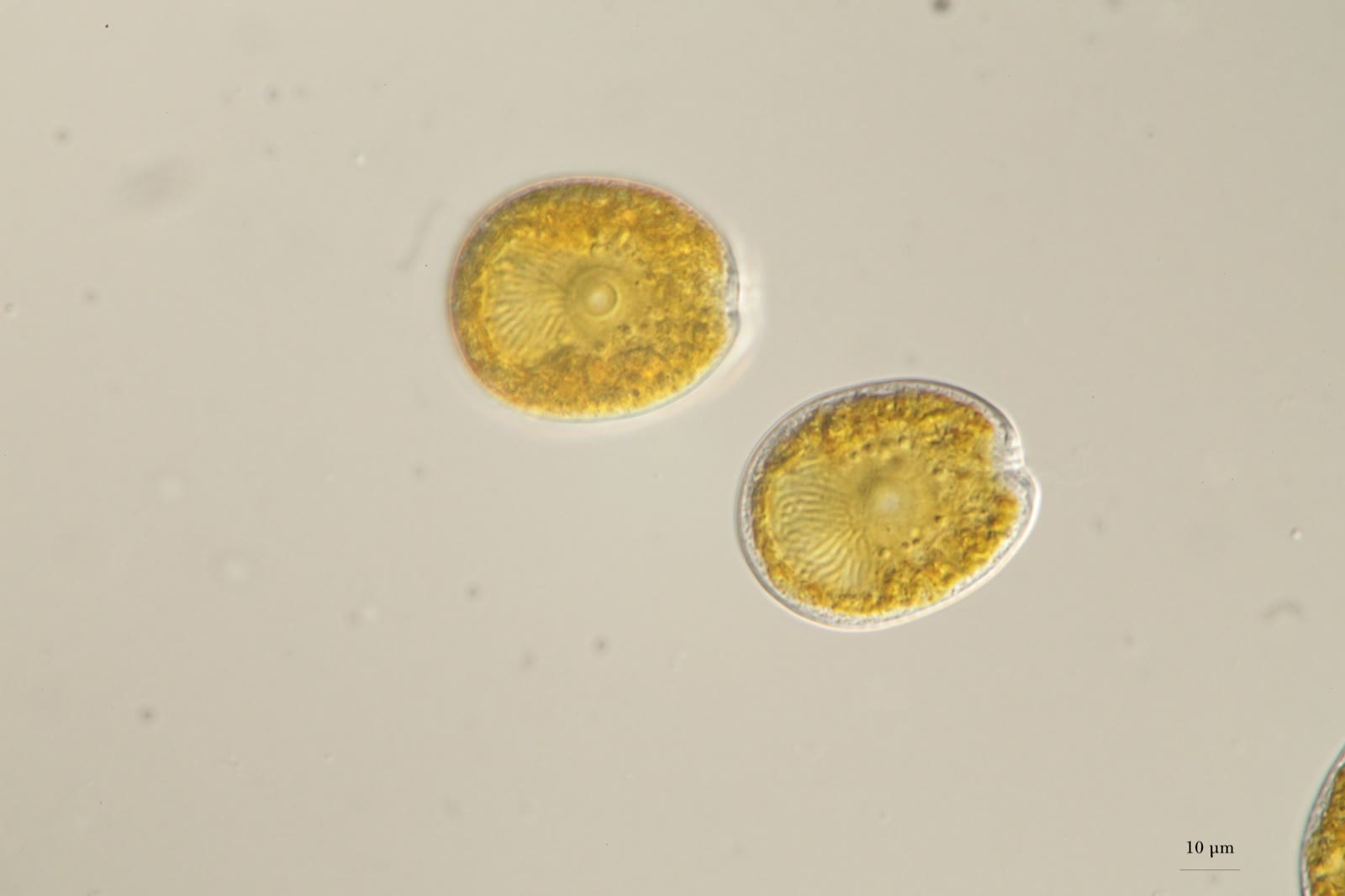
Pyrenoid. A large round body in the middle of the cell. Very obvious in Prorocentrum and in Amphidinium.
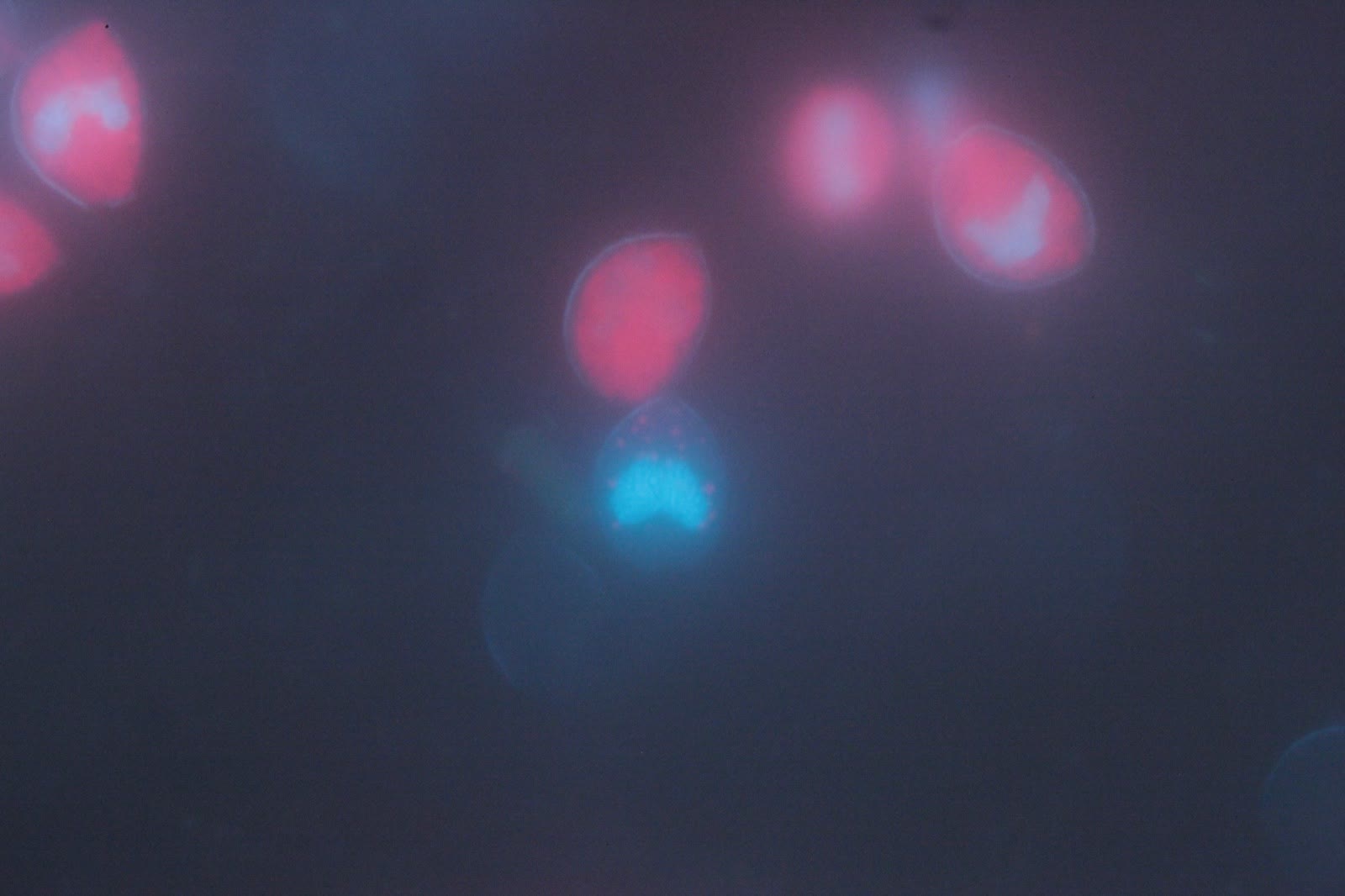
Nucleus (dinokaryon). Dinoflagellate nuclei are unusual: the chromosomes stay tightly condensed all the time. In a good light micrograph, as you focus up and down, this region looks cloudy and shows "spaghetti" or "brain" shapes. With a DAPI stain the nucleus lights up blue.
Chloroplast. Many dinoflagellates have a single chloroplast with many branches that fills the whole cell. That is what makes the amber lines you see in the cell.
Grooves. A typical dino has a groove running around the cell (the cingulum) and a groove running along the cell (the sulcus). The two different flagella sit in these grooves. In Amphidinium the cingulum is at the very top of the cell. Ostreopsis and Gambierdiscus are flattened from top to bottom like a disc, so when you look down at one, the cingulum runs around the outside edge.
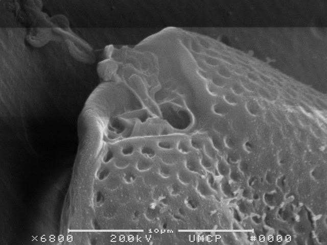
Plates (theca). Many dinoflagellates are wrapped in armor plates. Prorocentrum has two large halves called valves, and a notch at one end where the flagella emerge. Plate patterns are what most species names are based on, but you cannot see them well in a home microscope.
Flagella themselves will not be visible in a home microscope, so do not bother looking for them. Look at the shape and the motion.
Prorocentrum up close
Prorocentrum: mostly two big plates (valves). The grooves have shrunk into a notch at the apex, and there is no upper half.
Pictures of the parts
An undescribed Prorocentrum from a home aquarium (collected April 2012 by a marine aquarium hobbyist), 630X on a Zeiss Axioskop with a Canon EOS 5D Mark II. The cell rests on its left valve while the microscope is focused through the right valve, which is missing a notch at the apex. In Prorocentrum taxonomy the plates arranged in that notch matter, and the two different flagella emerge from pores there. The upper cell has the pyrenoid in focus; the lower cell has the dinokaryon in focus. The suture between the valves shows as a halo around the lower cell.Epifluorescence microscopy of Prorocentrum micans stained with DAPI. Nuclei appear blue from the DAPI; chloroplasts glow red from autofluorescence. The characteristic bi-lobed nucleus is visible.Scanning electron micrograph at 6800X showing the apical notch and the pores in the thecal plates of P. hoffmanianum. Scale bar 10 µm.
Confocal and 3D views
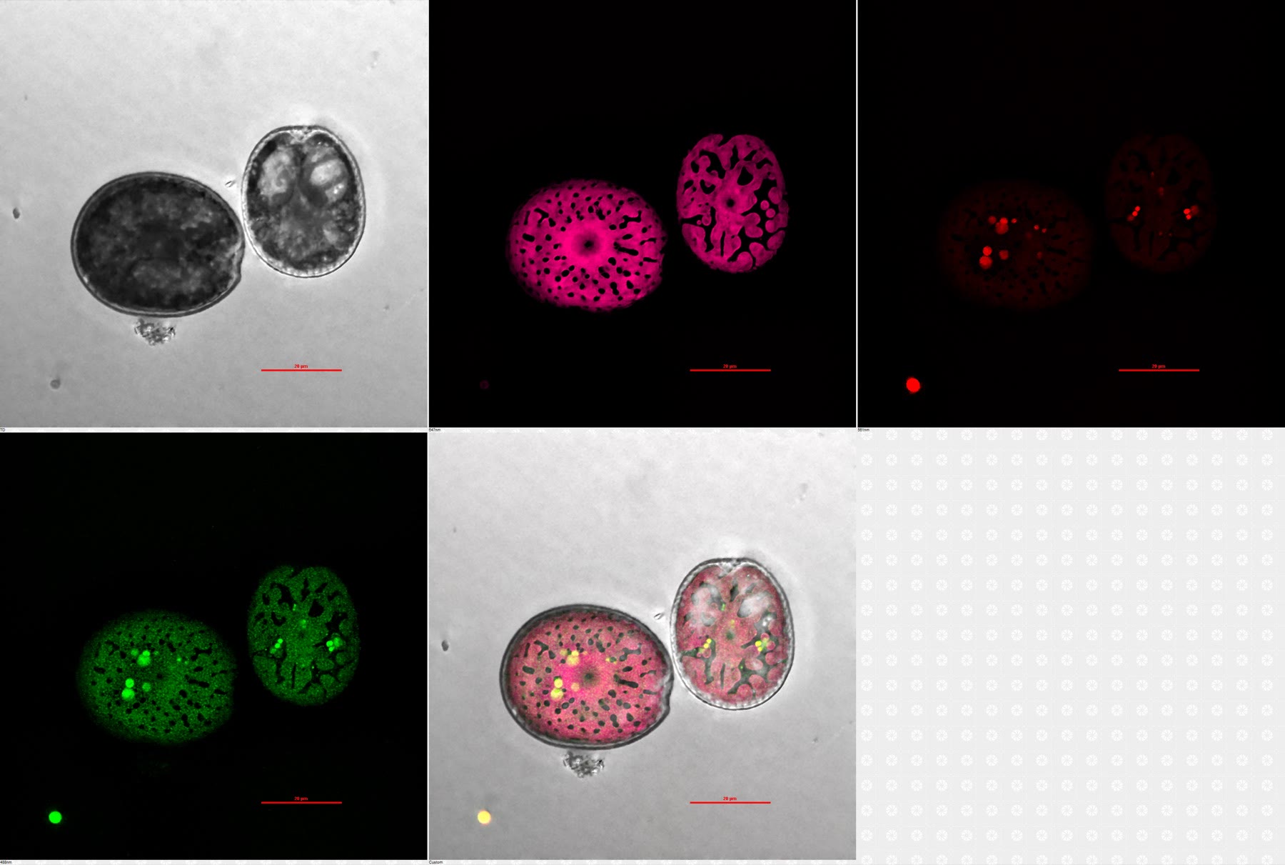
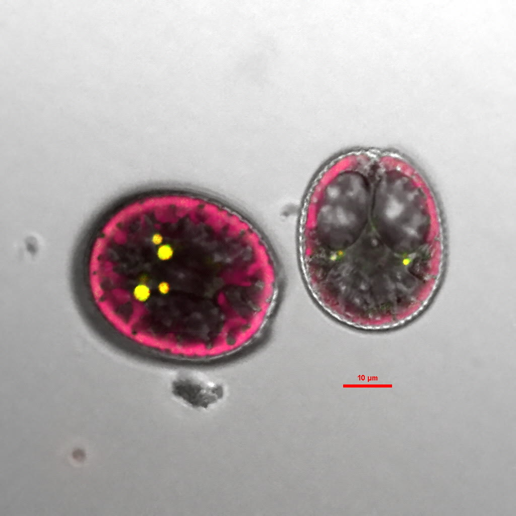
A hobbyist with access to a research microscope imaged one of these tank dinos with confocal microscopy and rendered the cells in three dimensions. These show the pores in the cell surface, the chloroplasts, and the overall shape in a way that regular microscopes cannot.
Note: the grayscale layers are DIC (differential interference contrast), which needs special optics. A standard home microscope will not show these fine details, so if you do not see these structures in your own sample, it could still be a dino.
Confocal microscopy of two dinoflagellate cells from a reef tank. Panels: DIC (a contrast technique that a standard home microscope cannot do), individual fluorescence channels, and an overlay. Pores are visible in the cell surface of the left cell. Image courtesy of a hobbyist.Overlay of DIC and fluorescence channels at 60X. Scale bar is 10 µm. The fine detail in the DIC layer needs specialized optics, so you will not see this in an ordinary microscope. Image courtesy of a hobbyist.3D rendering of the cell from a confocal Z-stack. Video courtesy of a hobbyist.3D rendering showing the theca (cell wall) of another cell, covered in pores. Video courtesy of a hobbyist.